


Question to the expert: Do I need to do an MRI just in case, and how does it threaten
Text: Gayana Demurina
RESPONSES TO THE MAJORITY OF US QUESTIONS we used to search online. In the new series of materials we ask such questions: burning, unexpected or widespread - to professionals in various fields.
Methods of examination, which allow you to see different organs and systems without pain and incisions, including even before the birth of a person, are called visualizing (or imaging techniques in English). True, many still doubt that these methods are safe: there are rumors about the dangers of even such an ordinary thing as ultrasound. As a result, two extremes arise: some fear visualization studies as fire, others insist on regular “tomography of everything”. How grounded are the concerns? Who needs such research and when? Should they be afraid of pregnant? We asked an expert to answer these questions.
Sergey Morozov
Chief Freelance Radiation Diagnostic Specialist, Moscow Department of Public Health
 Experiences about the safety of hardware examinations are quite understandable, because they somehow affect the cells of the body. The first thing we think about is how it will affect health in the future (especially if the word “radiation” sounds in the sentence). But in fact, not all types of imaging diagnostics use radiation: ultrasound and MRI have nothing to do with it.
In the case of ultrasound, the device creates oscillations, or waves; when an ultrasonic wave reaches tissues with a certain acoustic impedance, it is refracted. The part of the wave that acts on the tissue with less resistance will be absorbed by them and will pass on, and the other part, before which the resistance of the tissue is stronger, will be reflected. Roughly speaking, the more ultrasonic waves are reflected, the brighter and clearer the picture will be on the screen of the device. With MRI a slightly different story - but the main role here also belongs to the waves, only electromagnetic. They create a strong magnetic field and fix the response to it from some particles (the nuclei of hydrogen atoms are responsible for this). In fact, the device registers the response of the electromagnetic radiation of the body and displays the image. This is not a “photo” of the organ under study, but rather a map of its electromagnetic signals.
Such methods are safe for the patient's health, since they propagate sound or electromagnetic waves that cannot change the cell structure. Ionizing radiation (for example, x-rays or gamma rays, which are used by computed tomography) acts differently: the wavelength with such an effect can turn neutral particles in our tissues into charged, that is, ions (hence the name). For health, it is dangerous because the structure of tissues is changing. If the ionization catches the dividing cells by surprise and affects the protein synthesized using DNA, the resulting anomaly will repeat itself many times, like on a conveyor. So there are mutations that can lead, for example, to cancer.
Of course, this is not a reason to categorically refuse x-rays or CT. The point is the dose of radiation; for structural changes to start, it must be very large (symptoms of acute radiation sickness manifest themselves at a radiation level of 300 millisievert, and the safe dose is up to 100 millisievert). Modern diagnostic devices in this regard spare the body: for example, during lung X-rays, the patient may receive less than 1 mSv of radiation, with CT, the figures will vary depending on the area being studied, but in general should not exceed 16 mSv. At higher doses, radiation treats cancer - this is called radiation therapy. At the same time, the risk of developing a second tumor is not excluded, although this happens very rarely.
It turns out that to achieve a dangerous dose of radiation is difficult, and you should not be afraid of surveys. First, the harmful effects of ionizing radiation have so far been fixed only within the framework of large catastrophes, as in Chernobyl, where radiation doses were incredibly high. Secondly, we receive a certain proportion of radiation without medical examinations: a person who regularly leaves home receives up to 2-3 mSv of radiation per year. Our body has adapted to this kind of load and copes with it using protective mechanisms, including immune cells that capture and destroy cells with abnormalities, as well as apoptosis (programmed cell death).

Use only safe methods, so as not to encounter radiation at all, rather utopia than reality
On the other hand, it is not worth doing radiation diagnostics in any incomprehensible situation: although the harm of radiation in small doses remains in question, experts try not to expose patients to radiation in vain. Some organs are especially sensitive to radiation - these are the thyroid gland, the skin, the retina, the glands (including the milk), the organs of the small pelvis. In order to protect patients, certain protocols are followed: for example, lead aprons blocking X-rays are used, and the devices are adjusted so that a minimum dose sufficient for obtaining a good image is used.
Experts treat children and pregnant women with extreme caution: if the examination is recommended, but there is no urgent need for it, it may be postponed for some time. On the other hand, radiography of the teeth is safe for pregnant women, if done according to all the rules, the source of infection in the mouth, that is, caries or pulpitis, is much more dangerous for both mother and fetus. Ultrasound and MRI during pregnancy can be done without fear - with ultrasound used to determine not only the sex of the child, but also the risk of developing Down syndrome or congenital anomalies. The dangerous effect of ultrasound and MRI on the fetus is no more than a harmful myth, because there is no ionizing radiation from such studies.
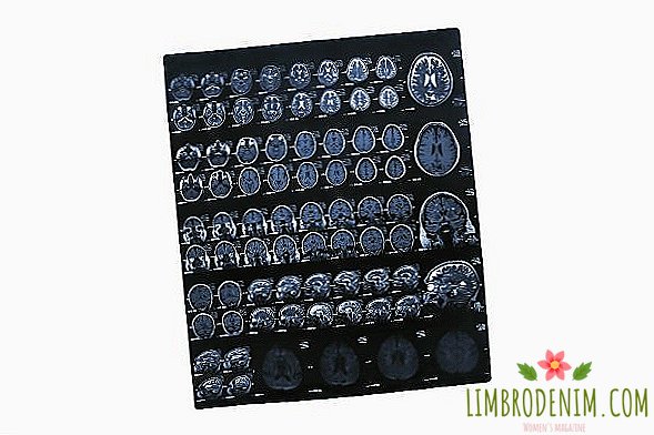
Use only safe methods, so as not to encounter radiation at all, rather a utopia than reality. If only because different types of diagnostics allow to look at the study area in different ways. The mechanisms of CT and MRI do not coincide, but they have one task - to display the object in three-dimensional form. With the help of computed tomography, fractures, hemorrhages, vascular function, and the state of the abdominal cavity are better diagnosed, although in general this method is also suitable for other cases. MRI is better suited for soft tissue, allows you to see the tumor and study, for example, the brain and spinal cord, although again this method can be used for other parts of the body.
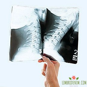
Ultrasound, on the contrary, has a limited spectrum of action. It is believed that it does not see the organs that are hidden behind the bones (the ultrasonic wave just does not reach them). And yet it is not amenable to automation, that is, a specialist is needed to interpret the ultrasound results. Nevertheless, it is easy to install the device right at the patient’s bed, which you cannot do, for example, with a massive MRI tunnel. Classical X-ray diagnostics are now being used less frequently than before, but sometimes it is impossible to do without it, for example, before complex operations. In fact, much depends not only on the purpose of the study, but also on the price, time and, in fact, the presence of the device in the clinic.
A healthy person under the age of forty does not need to undergo regular tomography. It should be recorded to the doctor when something really bothers. If it seems that you need something like a medical examination, it is enough to go through a simple check-up program (it usually includes ultrasound of various organs, ECG and echocardiography — an ultrasound of the heart, but it may also include a chest radiograph). Older people have x-ray examinations shown in regular examinations. For example, after fifty or sixty years, annual lung cancer screening is recommended - that is, CT scan of the lung, and after forty, women are also given breast cancer using mammography.
Photo: dmitrysteshenko - stock.adobe.com, Mandrixta - stock.adobe.com